
Abstract
In italicized text, there are instructions and descriptions for what to write in this section. Please delete these sections once you have finished writing your portion of the template.
If you have not finished this section yet, do not delete this section, and submit your paper with instructions undeleted.
The abstract gives the reader a 1-2 sentence summary of current research on your topic, a 1-2 sentence summary of the downfalls of the current research methods, and 1-2 summary of your proposed experiment. Your proposal includes the research method name and the targeted subjects/research object/community. The abstract ends with a sentence telling the reader how your experiment will benefit current research.
This template was based on the World Health Organization’s 2000 Guidelines for Research Proposals.
UNDP/UNFPA/WHO/World Bank Special Programme of Research, Development and Research Training in Human Reproduction. (2000). Preparing a research project proposal : guidelines and forms, 3rd ed. World Health Organization. https://apps.who.int/iris/handle/10665/66685
-
Introduction
Literature Review
Cancer development in the body occurs when a harmful mutation in a somatic cell causes rapid, uncontrolled proliferation, leading to chunks of malignant cells that create tumors. One way a cell may undergo a harmful mutation is through a genetic alteration of its DNA, which some viruses develop the ability to disrupt translations through viral replication. As the virus enters a cell, it can force the cell to translate its own genetic code, which in the process can create proteins that can switch on or off cell division regulators in a cell, leading to potential malignancy if left unattended. Both Epstein-Barr virus (EBV) and Human T-lymphotropic virus type-1 (HTLV-1) are some of the earliest examples of oncogenic viruses that possess this tumor-developing capability, leading to Burkitt Lymphoma (BL) and Adult T-cell leukemia/lymphoma (ATL) respectively. However, both are vastly different, as EBV is classified as a double stranded DNA (dsDNA) herpesvirus and HTLV-1 is a positive-strand RNA (+ssRNA) retrovirus.
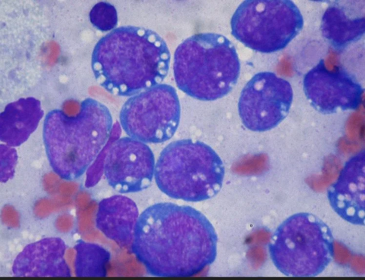
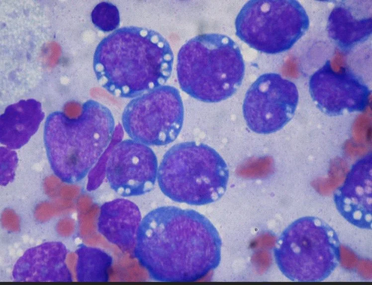
Burkitt Lymphoma was first discovered by British surgeon Denis Burkitt in 1958, when he noticed many of the children in Uganda that had tumors had a previous history with malaria and encounters with the Epstein-Barr virus. The virus is known to cause mononucleosis, otherwise known as mono or kissing disease, but it can also infect patients with various lymphomas and nasopharyngeal carcinoma, most notably BL, which can lead to patients experiencing tumor growth in the mouth and abdominal regions, along with vomiting, abdominal pain, and nausea. HTLV-1 was first found in Japan in 1979 by multiple American and Japanese doctors and scientists3, after several cases where patients with ATL had traces of HTLV-1 antibodies in their immune system. ATL causes hypercalcemia, swelling pain in the liver and spleen, and sometimes skin rashes. EBV and BL have some connection with the surface glycoprotein expressed on EBV, gp350/220, performing endocytosis into a B-lymphocyte via the CD21 receptor(Young), while HTLV-1 specifically targets heparan sulfate proteoglycans (HSPG) for entry, among other receptors found on the T-lymphocytes()Jones KS). There are no preventative measures yet, but research indicates that potential recombinant vaccines may be developed to help create antibodies for immunity, or specific chemotherapies that can be administered to shut down key steps in its viral replication.
Rationale
The Epstein-Barr virus may have intensive amounts of research ever since its discovery, but to this day no approved vaccination is on the market (although several candidates have already been developed), and treatment of the lymphoma can be tedious if treatment is delayed. HTLV-1 associated diseases are difficult to treat, as it is a relatively new virus with similar viral composition as HIV. However, these viruses are not completely untreatable. Both retroviruses and herpesviruses contain a key distinctive feature, that being their spherical morphology and surface glycoprotein, which can be targeted and neutralized through vaccination development. As the COVID-19 pandemic continues to rage, protection through mRNA vaccines are providing countless people with immunity, which can be replicated with other dangerous viruses that have similar glycoprotein compositions. The reason why mRNA vaccinations should be tested is because this new technology can potentially pave a future to safer but effective vaccines, as live attenuated vaccines insert the actual virus (although at lesser amounts so a major immune response is not triggered), causing some immunodeficient patients to seek alternatives to dangerous viruses, including herpesviruses and retroviruses.
With the concept of antiviral chemotherapy, lymphomas may be treated or prevented by blocking specific parts of the viral replication process, which in turn can lead to cancerous cell prevention. Additionally, treating the lymphoma directly with radiochemotherapy can be used to eliminate the tumor before metastasis. Overall, the treatment for EBV and HTLV-1 and their associated diseases fall on 2 main potential therapies: mRNA vaccines and radiochemotherapy, and further research into finding such a treatment should be considered.
Objectives
The objective is to test out a possible mRNA therapy for EBV associated BL and HTLV-1 associated ATL. This will require 2 separate experiments, one for each virus and their respective diseases. For the EBV-associated BL, an mRNA strand enclosed in a temporary synthetic nucleocapsid with the genetic sequence to imitate the gp350/220 glycoprotein expressed on the virus will be inserted intravenously, and the B-lymphocyte will replicate and express the protein on its membrane. For the HTLV-1 associated ATL, a similar mRNA strand enclosed in a temporary synthetic nucleocapsid with the genetic sequence to imitate the gp46 glycoprotein will also be inserted intravenously, only this time that the T-lymphocyte will replicate and express the protein on its membrane. With the surface protein replicated, the immune system will launch a response and collect information to produce antibodies that will neutralize EBV and HTLV-1, effectively also inhibiting any tumor development associated with the viruses.
-
Design & Methodology
Design
Patients over the age of 18 and below the age of 65 are eligible to participate in clinical trials, with an emphasis on patients with high immunity to minimize risks. EBV surface glycoproteins gp350 and gp220 will be key targets of interest for mRNA vaccine development, and samples can be collected from cell cultures with the EBV present (may it be from a patient’s blood or artificially grown viruses). The procedure then becomes using PCR to amplify the genome responsible for creating only the glycoprotein, creating a strand of mRNA that will cause the immune cell to read and express on its surface, and minimizing the risk of launching a severe immune response, with minimal discomfort to the patient. HTLV-1 will follow closely, with research targeting specifically the gp46 glycoprotein, deducing the genetic sequence that codes for the specific protein, and creating an mRNA vaccine similarly to the potential EBV vaccine. Patients with immunity will be deliberately infected with the respective virus, and treatment and medication will be provided at no cost to all patients willing to take the risk. Data on antibody expression, treatment efficacy, and other patient information will be taken into consideration when completing analysis.
Multiple chemotherapeutic drugs can be administered to hopefully prevent the virus from continuing its replication process. In the process of viral replication, several key steps can be blocked off in hopes of eliminating the virus before exocytosis. Some specific targeted processes include preventing the cell from translating the virus RNA, preventing the cell from continuing protein synthesis, and preventing the viral assembly to form a mature virus cell. However, more research will be needed to determine if chemotherapy can be a resolute answer to the virus, as it has many mild to severe aftermaths depending on the patient.
Duration and Location
There is no set time or location to conduct the experiment as of yet.
Subjects
The target group, as briefly stated above, include volunteers that are over the age of 18 and under the age of 65, preferably those with high immunity. In addition, subjects of the experiment should avoid participating if they are currently suffering from HIV/AIDS or any other immunocompromising illness, or care for any people that are immunocompromised, or have any known medical problems such as allergic reactions that prevent them from taking vaccinations.
Procedures
Protein Purification and Isolation
This protocol is adapted from Lodish, H. (1970, January 01). Purifying, Detecting, and Characterizing Proteins. This method will be used to purify a protein and isolate any components to the target protein. This method will work for both EBV and HTLV-1.
-
Use nonionic detergents (Octylglucoside) in high concentrations to first break down the components of the virus.
-
Separate the particles using a centrifuge and perform differential centrifugation to obtain the glycoprotein. It should have higher mass and density due to its composition as a protein and a carbohydrate.
-
Identify the correct glycoprotein via NMR (nuclear magnetic reaction) spectroscopy. Steps to perform an NMR spectroscopy is found below: https://www.cif.iastate.edu/nmr/nmr-tutorials/sample-preparation
-
Use the proper amount of material - typical 1H NMR spectra require 5-25 mg of material.
-
Use the proper solvent and amount of solvent - Deuterated solvents including chloroform-D, acetone-D6, benzene-D6, deuterium oxide (D2O), DMSO-D6, ethanol-D6, and methanol-D4 can be used.
-
Prepare a sample in a secondary vial - transfer to an NMR tube with a glass Pasteur pipette.
-
Insert the sample into a Thin-Walled Economy NMR Sample Tube, 5mm, 500 MHz (Part Number 82005-328) NMR tube with the AVIII 600 MHz NMR Spectrometer. Avoid disposable NMR tubes in case of breakage.
-
Use an internal standard - Add a drop of TMS to 5-10 mL of deuterated solvent.
-
-
Once the identification is complete and the desired surface protein is obtained, identify the concentration for the respective protein and proceed onto PCR.
Polymerase Chain Reaction (EBV)
This protocol is adapted from Germi, R., Morand, P., Brengel-Pesce, K., Fafi-Kremer, S., Genoulaz, O., Ginevra, C., . . . Seigneurin, J. (2004, October 1). Quantification of gp350/220 Epstein-Barr Virus (EBV) mRNA by Real-Time Reverse Transcription-PCR in EBV-Associated Diseases. Retrieved July 28, 2021, from https://academic.oup.com/clinchem/article/50/10/1814/5640030. The process details the reverse-transcription PCR needed to obtain the RNA of the EBV:
In this study, we developed a method for quantifying a late transcript of the highly conserved EBV envelope glycoprotein gene, gp350/220 (12)(13), that uses real-time reverse transcription-PCR (RT-PCR), the Taqman® technology, and serial dilutions of in vitro transcripts to quantify mRNA. gp350/220 mRNA was quantified in EBV-infected cell lines and was then applied to a series of clinical samples.
Total RNA from cell lines (500 000 cells), clinical samples, or in vitro transcripts (5 μL) was extracted with the High-Pure-RNA-Isolation-Kit® (Roche), and residual DNA was removed by two 20-min incubations with DNase I. The RNA concentration was estimated spectrophotometrically and converted to copy number for the in vitro transcript.
The RT-PCR mixture (20 μL) contained 100 ng of total RNA from cell lines or 250 ng of total RNA from clinical samples, 3.25 mM manganese acetate (LightCycler-RNA-Master-Hybridization-Probes-Kit®; Roche), 0.3 μM upstream primer [gpU2; 5′-AGAATCTGGGCTGGGACGTT-3′; position 89585 ], 0.3 μM downstream primer (gpL2; 5′-ACATGGAGCCCGGACAAGT-3′; position 89784), and 0.3 μM probe [EBVGPq; 5′-(6-FAM)AGCCCACCACAGATTACGGCGGT(TAMRA)(Phosphate)-3′, where 6-FAM is 6-carboxyfluorescein and TAMRA is 6-carboxytetramethylrhodamine; position 89761]. The RT-PCR program (61 °C for 30 min, 3 min at 95 °C, and 40 cycles of 10 s at 95 °C, and 90 s at 60 °C), produced an amplified fragment of 200 bp.
Absolute quantification was performed with an external RNA calibration curve. The RNA calibrator was obtained by in vitro transcription of 1 μg of a constructed plasmid for 2 h at 37 °C (SP6/T7-Transcription-Kit®; Roche). The plasmid was obtained by cloning in pSPT18 the 200-bp gpU2/gpL2 RT-PCR product, obtained from B95-8 cell line RNA. Serial dilutions of the RNA calibrator were analyzed by real-time RT-PCR, and a wide linear range (50 to 5 × 10⁶ copies) was obtained, indicating the efficiency of the amplification and the sensitivity of the method.
The amplification efficiency in gp350/220-negative samples was controlled by real-time RT-PCR of a reference gene using LightCycler-h-PBGD-Housekeeping-Gene-Set® (Roche). The specificity of the method was confirmed by use of EBV-negative BJAB cells and cells infected by five other herpesvirus strains. A PCR without the reverse transcription step confirmed the absence of DNA contamination.
Polymerase Chain Reaction (HTLV-1)
This protocol is adapted from Maria de Nazaré do Socorro de Almeida Viana, Nobre, A. F., Jr, E. C., Silva, I. C., Pinheiro, B. T., Pereira, C. C., . . . Sousa, M. S. (2018, May 02). Stability of the HTLV-1 glycoprotein 46 (gp46) gene in an endemic region of the Brazilian Amazon and the presence of a significant mutation (N93D) in symptomatic patients. Retrieved July 27, 2021, from https://virologyj.biomedcentral.com/articles/10.1186/s12985-018-0984-9. The chain reaction to copy the gp46 glycoprotein of the HTLV-1 virus is as detailed:
Following the genotyping of the HTLV, and the identification of the infected patients, the complete sequence of the gene for the HTLV-1 p46 glycoprotein was amplified. This was based on three reactions — (i) a PCR with the gp46F1/gp46R1 nucleotide primers, which generated a fragment of 1047 bps, (ii) a semi-nested PCR using the gp46F1/gp46R2 primers, which produced a fragment of 749 bps , and (iii) a semi-nested PCR using the gp46F2/gp46R1 primers, which generated a fragment of 627 bps, which complements the fragment amplified in the preceding step (Supplementary Scheme 1).
The PCR for the complete fragment of the gp46 gene was run in 12.5 μL of Go Taq (2×) Green Master Mix, 8.5 μL of water, 10 pmol (0.5 μL) of each primer (gp46F1: 5’CGCCGATCCCAAAGAAAAA3’ and gp46R1: 5’ACATGGAGCCGGTAATCCC3’) and 3 μL (100 ng) of DNA, with a final volume of 25 μL. In the first semi-nested PCR, the same quantity of Go Taq (2×) Green Master Mix was used, together with 10.5 μL of water 10 pmol (0.5 μL) of each primer, that is, gp46F1 and gp46R2 (5’GACGTGCCAAGTGGATAGGC3’), and 1 μL of the amplified DNA, with a final volume of 25 μL. The same amplification conditions were applied, to generate a fragment of 749 bps. In the second semi-nested PCR, 14.0 μL of Go Taq (2×) Green Master Mix was added to 9.2 μL of water, 10 pmol (0.4 μL) of each primer, gp46F2 (5’GCCCCTACTGGAAATTTCAGC3’) and gp46R1, and 1.0 μL of the DNA, for a final volume of 25 μL. A fragment of 627 bps was obtained using the same amplification conditions.
The amplification protocol for all the reactions was based on 30 cycles of 30 s at 94 °C for denaturation, 30 s at 55 °C for annealing, and 30 s at 72 °C for extension, with a final extension of 10 min at 72 °C, and then 10 min at 10 °C. The PCR products (6 μL) were electrophoresed at 50 mV for 1 h in 1.5% agarose gel.
Ex Vivo Testing for B-cells (EBV and BL)
This protocol is adapted from Selkirk, S M. “Gene Therapy in Clinical Medicine.” Postgraduate Medical Journal, The Fellowship of Postgraduate Medicine, 1 Oct. 2004, pmj.bmj.com/content/80/948/560. The process involves exposing B-lymphocytes directly from bone marrow cells to a dosage of the EBV mRNA vaccine, selecting cells that are capable of expressing the glycoprotein to reinsert for immune response, and testing the efficacy of the therapy by exposure to the EBV in small amounts.
-
Identify a surrogate cell population that has a high expression of CD21 receptors. Specifically, target B-lymphocytes produced by a patient’s bone marrow. Alternatively, spleen and lymph nodes have matured B-lymphocytes and can be points of interest for exposure to the EBV mRNA vaccine. Obtain additional samples in case of failure.
-
Administer a 0.3 mL dose of the EBV mRNA vaccine. If necessary, adjust the dosage amount to cell culture. Wait approximately 24 hours for cells to replicate the protein and select cells that have expressed the gp350/220 glycoprotein. Insert the cell culture back into the patient and wait 14 days for the immune system to react to the foreign antigen and create antibodies.
Additional note: ex vivo trials may be done with brown rats, also known as Rattus norvegicus. Rat bone marrow cells and rat spleen B-lymphocytes are preferred.
-
Once antibody levels are high, introduce the Epstein-Barr virus and test for efficacy. Wait 1-2 days and record virus and antibody levels. Wait 5 days and record virus and antibody levels again. Continue the process over the span of days and weeks until the virus is completely eliminated and/or the antibody levels grant enough immunity.
Ex Vivo Testing for T-cells (HTLV-1 and ATL)
This protocol is adapted from Selkirk, S M. “Gene Therapy in Clinical Medicine.” Postgraduate Medical Journal, The Fellowship of Postgraduate Medicine, 1 Oct. 2004, pmj.bmj.com/content/80/948/560. The process, very similar to EBV and BL, involves exposing T-lymphocytes directly from the thymus to a dosage of the HTLV-1 mRNA vaccine, selecting cells that are capable of expressing the glycoprotein to reinsert for immune response, and testing the efficacy by exposure to the HTLV-1 in small amounts.
-
Identify a surrogate cell population that has a high expression of T-cell receptors (including HSPGs). Specifically, target T-lymphocytes produced by a patient’s bone marrow or mature T-lymphocytes in the thymus. Obtain additional samples in case of failure.
-
Administer a 0.3 mL dose of the HTLV-1 mRNA vaccine. If necessary, adjust the dosage amount to cell culture. Wait approximately 24 hours for cells to replicate the protein and select cells that have expressed the gp46 glycoprotein. Insert the cell culture back into the patient and wait 14 days for the immune system to react to the foreign antigen and create antibodies.
Additional note: because of the malignant oncogenic potential of HTLV-1, first trials are strictly prohibited to be tested on humans. Test subjects like Rattus norvegicus are preferred for experimentation.
-
Once antibody levels are high, introduce HTLV-1 and test for efficacy. Wait 1-2 days and record virus and antibody levels. Wait 5 days and record virus and antibody levels again. Continue the process over the span of days and weeks until the virus is completely eliminated and/or the antibody levels grant enough immunity.
Until reports are written for both tests and approval is given by the FDA, do not proceed on to human clinical trials.
mRNA vaccinations (EBV and HTLV-1) - Human Clinical Trials
-
Inject mRNA vaccine intravenously into patients and have them wait for 30 minutes. Observe any abnormal immune responses and treat accordingly. Injection process is very similar to administering the COVID-19 vaccine. Adapted from CDC, “Administration Overview for Pfizer-BioNTech COVID-19 Vaccine.” Centers for Disease Control and Prevention, Centers for Disease Control and Prevention, 21 May 2021, www.cdc.gov/vaccines/covid-19/info-by-product/pfizer/index.html.
-
Follow aseptic technique. Perform hand hygiene before vaccine preparation, between patients, when changing gloves (if worn), and any time hands become soiled.
-
Remove vaccine from the freezer or refrigerator. Allow vaccine to come to room temperature. Vials can be held at room temperature for up to 2 hours before mixing. After 2 hours, return unmixed vials to the refrigerator.
-
Before mixing, check the expiration dates of the vaccine and diluent. NEVER use expired vaccine or diluent.
-
With the vaccine at room temperature, gently invert the vial 10 times. Do NOT shake the vial. If the vial is shaken, contact the manufacturer. The vaccine is white to off-white in color and may contain opaque particles. Do not use if liquid is discolored.
-
Using a new, sterile alcohol prep pad for each vial, wipe off the stoppers of the diluent and vaccine vials.
-
Using a 21-gauge (or narrower) needle, withdraw 1.8 mL of 0.9% sodium chloride (normal saline, preservative-free) into a mixing syringe. After use, discard diluent vial and any remaining diluent.
-
Do NOT use or save the remaining vaccine diluent to mix additional vaccine or for other uses.
-
Do NOT use bacteriostatic normal saline or other diluents to mix the vaccine.
-
-
Inject 1.8 mL 0.9% sodium chloride (normal saline, preservative-free) diluent into the vaccine vial.
-
Using the mixing syringe, remove 1.8 mL of air from the vaccine vial to equalize the pressure in the vaccine vial.
-
Gently invert the vial containing vaccine and diluent 10 times. The vaccine will be off-white in color. Do not use if discolored or contains particulate matter. Do NOT shake. If the vial is shaken, contact the manufacturer.
-
Note the date and time the vaccine was mixed on the vial.
-
Keep mixed vaccine between 2⁰C and 25⁰C (36⁰F and 77⁰F) and administer within 6 hours. Discard any unused vaccine after 6 hours. Do not return to freezer storage.
-
Minimize exposure to room light, and avoid exposure to direct sunlight and ultraviolet light.
-
-
Choose the correct equipment, including the correct needle size.
-
Use a new, sterile needle and syringe for each injection.
-
Low dead-volume syringes/needles can be used to extract 6 doses from a vial. If all low dead-volume syringes are not available use a combination of low dead-volume syringes and non-low dead-volume syringes.
-
-
Cleanse the stopper on the vial of mixed vaccine with a new, sterile alcohol prep pad. Withdraw 0.3 mL of mixed vaccine into the syringe. Ensure the prepared syringe is not cold to the touch.
-
Regardless of the type of syringe used, ensure the amount of vaccine in the syringe is 0.3mL.
-
If the amount of vaccine remaining in the vial cannot provide a full 0.3mL dose discard the vial and contents.
-
Do not combine vaccine from multiple vials to obtain a dose.
-
-
Remove any significant air bubbles with the needle still in the vial to avoid loss of vaccine. Use the same needle to withdraw and administer the vaccine, unless contaminated or damaged.
-
-
Have patients come back after 2 weeks to take a sample of their blood to test for antibody levels, and introduce the virus to the patients and record the antibody levels after vaccination. Report any problems and steps to improve.
III. Discussion
Expected Problems
One possible outcome of the vaccine is an immune system overreaction, which may occur due to the presence of an unknown antigen. As such, inflammatory syndromes ranging from light fevers to severe Multi-Inflammatory Syndrome (MIS) can develop, potentially 1-2 days after the therapy, albeit rarely, and other symptoms like gastrointestinal pain or discomfort may occur. Quick solutions to the problem, however, include antihistamine medication including acetaminophen (Tylenol) for fever relief and temporary pain relief. Additionally, if the dosage is insufficient or the immune system does not react to the foreign antigen at all (in which the data reveals no antibodies are expressed in a particular patient), the process should be reviewed for any flaws in the PCR or replication process.
Another problem that may occur includes specific mutations that may affect the development of mRNA in the virus or the glycoprotein itself. As viruses are commonly adapting to different environments by evolving, both EBV and HTLV-1 may encounter mutations in their glycoprotein or genetic code to attach to a host quicker or more efficiently, leading to decreased efficacy or even reinfection after full immunity. Generally, these vaccines should offer protection against the original and herd immunity can stop it before it evolves any further, but should the occasion arise, booster shots with the updated mRNA mutations should be processed with the same procedures as before.
Expected Outcomes
Data on the vaccine efficacy will be collected onto a table denoting specifically antibody levels. Any correlation with the drug dose amount with the antibody level should be noted. Additionally, if any tumor development or allergic reactions occurs, the data will be recorded and the patient will be sent immediately to be diagnosed and treated accordingly. At the current standpoint, the experiment consists of an mRNA strand that codes for a specific surface glycoprotein expressed on two oncoviruses, Epstein-Barr virus and Human T-lymphotropic virus, triggering the immune system to create antibodies against future infections. Those specific glycoproteins, gp350/220 for EBV and gp46 for HTLV-1, will be integral catalysts to increase immunity to dangerous viruses, as well as pave the way for future vaccine development against viruses with oncogenic potential.
IV. Ethical Considerations
Informed Decision Making and Confidentiality Consent Forms
Because the experiment involves exposing viruses and potentially cancer to patients, the procedures should be heavily restricted and patients participating in the study should be notified beforehand about the risks, especially during human clinical trials. The subject should always know that a compensation (undetermined amount at the moment) as well as treatment is provided if consent is given, and that all experimental therapies given throughout the trials are tested and proven to be safe for human consumption.
Risk-Benefit Assessment
The risks in taking part of the study, as listed before, include exposure to the Epstein-Barr virus or Human T-lymphotropic virus type-1 and all of their associated diseases (including, but not limited to, infectious mononucleosis, nasopharyngeal carcinoma, and EBV-associated Burkitt’s Lymphoma for EBV and Adult T-cell leukemia/lymphoma for HTLV-1). In the worst case scenario, the treatment is unresponsive when exposed to the virus, and the patient develops cancer. However, all patients that participate in the experiment will be closely monitored for any developments and immune response, and will not be denied treatment for any reason whatsoever, so the potential outcome of a dead patient is extremely low.
If the treatment is a success, the vaccine will grant the person protection against EBV and/or HTLV-1 and all associated diseases, including the cancers caused by the viruses. Additionally, compensation is given to all patients regardless of outcome.
Participating in the research will not only help mitigate oncoviruses and their cancers, but also pave a way for the future of mRNA vaccination development as more research is performed on the potential of the vaccine. Insight to oncotherapy may also be discovered while undergoing the experiment, so patients that participate are taking part in a revolutionary study to take one step closer into finding a cure for cancer and helping patients that suffer from these deadly diseases worldwide.
Bibliography
-
“Administration Overview for Pfizer-BioNTech COVID-19 Vaccine.” Centers for Disease Control and Prevention, Centers for Disease Control and Prevention, 21 May 2021, www.cdc.gov/vaccines/covid-19/info-by-product/pfizer/index.html.
-
Chemotherapy. (2020, March 05). Retrieved July 27, 2021, from https://www.mayoclinic.org/tests-procedures/chemotherapy/about/pac-20385033
-
Coiffier, B., & Sarkozy, C. (2016, December 02). Diffuse large B-cell lymphoma: R-CHOP failure-what to do? Retrieved July 27, 2021, from https://www.ncbi.nlm.nih.gov/pmc/articles/PMC6142522/
-
Esau, D. (2017, September 25). Viral Causes of Lymphoma: The History of Epstein-Barr Virus and Human T-Lymphotropic Virus 1. Retrieved July 27, 2021, from https://www.ncbi.nlm.nih.gov/pmc/articles/PMC5621661/
-
Germi, R., Morand, P., Brengel-Pesce, K., Fafi-Kremer, S., Genoulaz, O., Ginevra, C., . . . Seigneurin, J. (2004, October 1). Quantification of gp350/220 Epstein-Barr Virus (EBV) mRNA by Real-Time Reverse Transcription-PCR in EBV-Associated Diseases. Retrieved July 28, 2021, from https://academic.oup.com/clinchem/article/50/10/1814/5640030
-
Jones, K. S., Lambert, S., Bouttier, M., Bénit, L., Ruscetti, F. W., Hermine, O., & Pique, C. (2011, June). Molecular aspects of HTLV-1 entry: Functional domains of the HTLV-1 surface subunit (SU) and their relationships to the entry receptors. Retrieved July 27, 2021, from https://www.ncbi.nlm.nih.gov/pmc/articles/PMC3185769/
-
Jones KS;Petrow-Sadowski C;Bertolette DC;Huang Y;Ruscetti FW;. (2005, October). Heparan sulfate proteoglycans mediate attachment and entry of human T-cell leukemia virus type 1 virions into CD4 T cells. Retrieved July 27, 2021, from https://pubmed.ncbi.nlm.nih.gov/16188972/
-
Lodish, H. (1970, January 01). Purifying, Detecting, and Characterizing Proteins. Retrieved July 27, 2021, from https://www.ncbi.nlm.nih.gov/books/NBK21589/
-
Maria de Nazaré do Socorro de Almeida Viana, Nobre, A. F., Jr, E. C., Silva, I. C., Pinheiro, B. T., Pereira, C. C., . . . Sousa, M. S. (2018, May 02). Stability of the HTLV-1 glycoprotein 46 (gp46) gene in an endemic region of the Brazilian Amazon and the presence of a significant mutation (N93D) in symptomatic patients. Retrieved July 27, 2021, from https://virologyj.biomedcentral.com/articles/10.1186/s12985-018-0984-9
-
Selkirk, S M. “Gene Therapy in Clinical Medicine.” Postgraduate Medical Journal, The Fellowship of Postgraduate Medicine, 1 Oct. 2004, pmj.bmj.com/content/80/948/560
-
Young KA;Herbert AP;Barlow PN;Holers VM;Hannan JP;. (2008, November). Molecular basis of the interaction between complement receptor type 2 (CR2/CD21) and Epstein-Barr virus glycoprotein gp350. Retrieved July 27, 2021, from https://pubmed.ncbi.nlm.nih.gov/18786993/
