
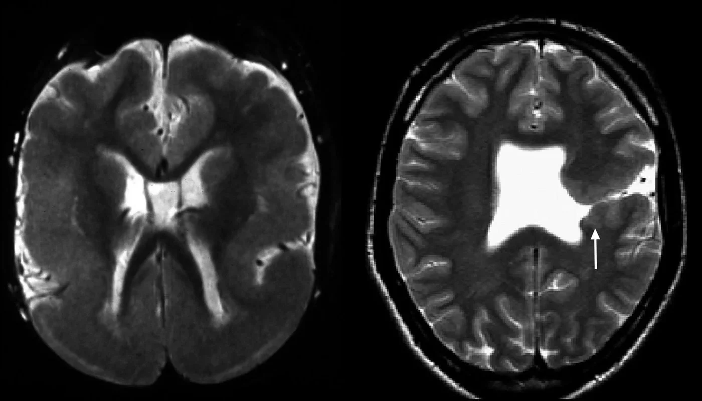
A team led by Dr. Martin Bax aimed to identify the correlates between different methods of identification for the disorder Cerebral Palsy. The paper took into account 585 children born with cerebral palsy between the years of 1996-1999, as the paper was published in 2006, the children were between the ages of 10 and 15. 431 of the children used in the experiment were recruited and assessed. Of the children in the study, 61.9% were male, and the rest were female. As assessment methods vary, the study’s objective was to identify correlates and comparisons between those assessed for CP via MRI brain scan and those assessed via clinical methods. Thus, the independent variable was the method of diagnosis/assessment for the disorder- clinical or MRI- and the dependent variable is the result- correlates and information found via both methods. The experiment required no control groups. The study took place in Europe, centering in major cities, including North West and North East London, England; Edinburgh, Scotland; Lisbon, Portugal; Dublin, Ireland; Stockholm, Sweden; Tübingen, Germany; and Helsinki, Finland.
Some important findings include the high rate of infections reported by mothers during pregnancy, with the rate being around 39.5%. In addition, 235 children or 54% of the total children were born at term while 47 children, 10.9%, were born preterm. They were either born 28 weeks early or less. Clinically, 26.2% of children had hemiplegia (paralysis on one side of the body), 34.4% had diplegia (paralysis of corresponding parts on both sides of the body. It typically affects the legs more severely than the arms), 18.6% had quadriplegia (paralysis from the neck down), 14.4% had dyskinesia (impairment of voluntary movement), 3.9% had ataxia (the loss of full control of bodily movements), and 2.6% had other types of cerebral palsy. Brain MRI scans showed that white-matter damage of immaturity, including periventricular leukomalacia (PVL), was the most common finding (42.5%), followed by basal ganglia lesions (12.8%), cortical/subcortical lesions (9.4%), malformations (9.1%), focal infarcts (7.4%), and miscellaneous lesions (7.1%). Only 11.7% of these children had normal MRI findings. Some advantages of this method was that it lets doctors identify preventive strategies for Cerebral Palsy and that the experiment took into account multiple factors. We can also tell that the experiment outcomes were precise because the experiment took place throughout 8 European study centers. These MRI findings suggest that obstetric mishaps might have occurred in a small proportion of children with CP.
APA Source Citation:
Bax, M., Tydeman, C., & Flodmark, O. (2006, October 2006 4). Clinical and MRI Correlates of Cerebral Palsy The European Cerebral Palsy Study. JAMA. Retrieved October 15, 2021, from https://jamanetwork.com/journals/jama/article-abstract/203508
