
Introduction
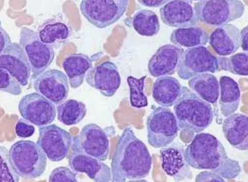
Cancer is a disease in which mutations within our own cells cause them to grow uncontrollably and metastasis in other parts of our body. There are many different subsections of cancer, the specific type of cancer is named after the origin. Oftentimes, most cancers form tumors which are a group of multiple cells that are mostly cancerous. One type of cancer that does not usually create tumors is leukemia, which is a cancer of the blood. This type of cancer often begins in white blood cells, but can also occur in other blood cell types. Leukemia is classified based on the speed of its growth, acute (fast) and chronic (slow). This paper will specifically focus on acute myelogenous leukemia. This type of cancer occurs when the bone marrow accumulates mutations and produces myeloblasts, immature white blood cells that become leukemic. It is called acute because of its rapid development.
Normal Blood Cell Formation
In a healthy body, blood cells are formed from the bone marrow, the soft inner part of the bones. In here there are blood stem cells that can develop into different types of blood cells, lymphocytes (B and T cells) or myeloid cells. Myeloid cells can further develop into other white blood cells, red blood cells, or platelets (fragments of cells that bind to broken blood vessels).
Juluri 2
The three types of white blood cells are monocytes - which develop into macrophages -, granulocytes, and lymphocytes.
Causes of AML
Chromosomal Abnormalities
In a normal healthy body, the above processes would occur without resulting in cancer. During the DNA replication process, errors that occur during division are normally corrected or the misformed cell is killed. It has been found that patients with AML often have changes in the c-KIT, FLT3, and RAS genes that prevent the bone marrow cells from properly developing or assist in their uncontrollable growth. The changes in these genes are more commonly chromosome changes rather than mutations in the DNA. Chromosome changes include translocation, deletions, inversions, and addition/duplication. A translocation is when one part of a chromosome breaks off and attaches to another gene, which can affect the function of the genes and possibly turn them into oncogenes. In AML, common genes that are turned off due to translocation are RARa and RUNX1, which control the maturation of normal blood cells. A deletion is when a part of a chromosome is cut off, making it dysfunctional. If the chromosome is a tumor suppressor, it would be unable to prevent the uncontrolled growth. An inversion is when one part of the gene gets flipped over resulting in the loss of the gene. An addition or duplication modification is when there is an extra part of a chromosome or an extra chromosome. If this happens to an oncogene, it will be a serious problem.
Signalling Pathways
It has also been found through the 2004 study conducted by Bernasconi et.al using animal models that just translocations and inversions are not enough to cause AML. Instead, there also have to be point mutations affecting the receptor tyrosine kinases (RTKs). These mutations are thought to help the cells with damaged chromosomes proliferate and grow uncontrollably.
Figure 1
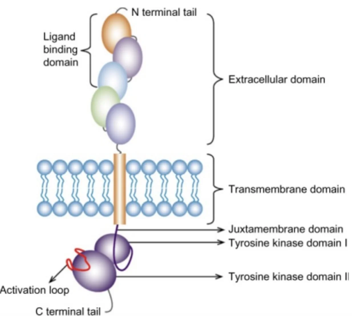
Note: This is an image of a Class III RTK, the most common gene in which mutations are found in AML patients.
Figure 2
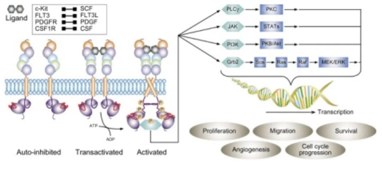
When RTK is activated, there will be constant phosphorylation of downstream effectors and signalling pathways that release biochemical signals that then affect Ras/MAPK, STAT, and PI3K, which are known to be the major oncogenic signals.
Risk Factors
AML is not typically caused by inherited genes but rather by mutations that occur during a person’s lifetime. While this disease can occur anytime, it is more common in those who are 65 and older. Additionally, for some unexplained reason men are more likely to be diagnosed with AML than women. AML has also been linked with exposure to radiation, chemicals such as benzene, and smoking. Oftentimes, people who have previously been treated with radiation for cancer treatment later develop AML. People who have other blood disorders or genetic disorders (Down Syndrome) also have a higher risk of developing AML. While AML is not genetic, people who have close family members that have been previously diagnosed have a higher risk.
Some other risk factors that have not been conclusively linked are: exposure to chemicals/solvents in the workplace, electromagnetic fields (power lines), and herbicides/pesticides.
Types of AML
There are many different types of AML. The two major systems used to classify AML were made by the WHO (World Health Organization) and the FAB (French American British) classifications. The major groups created by WHO are:
● AML with multilineage dysplasia
● therapy-related myeloid neoplasm
● AML that is not otherwise categorized
● Myeloid sarcoma
● Myeloid proliferations related to Down Syndrome
● Undifferentiated or biphenotypic acute leukemia
The following classification system was created by the FAB.
M0: Myeloblastic without differentiation
M1: Myeloblastic with little or no maturation
M2: Myeloblastic with maturation
M3: Promyelocytic
M4: Myelomonocytic
M4eo: Myelomonocytic with eosinophils
M5a: Monocytic without differentiation (monoblastic)
M5b: Monocytic with differentiation
M6: Erythroleukemia
M7: Megakaryocytic
AML is mostly classified based on what normal cells the cancerous cells resemble. The most common type of AML is myeloid leukemia which is a cancer in the cells that produce neutrophils. Another common type is monocytic leukemia, which is named after the cancer cells that look similar to monocytes. AML is also classified based on the chromosomal changes found in the cells. This can help the doctors determine the most effective treatment for the patients. By grouping using chromosomal changes, doctors can better identify what the survival rates are. For patients who have abnormalities in chromosome 16 at the p13 and q22 bands or translocation between chromosomes 8 and 21 have a more favorable outcome. Patients that have no chromosomal abnormalities or a translocation between chromosomes 9 and 11 have a less favorable outcome. Patients with an unfavorable outcome (low chances of completing curing the leukemia or treatment working) have deletions in chromosome 7 and 5, extra copies of chromosome 13 and 8, and complex changes on chromosomes.
Diagnosis
To diagnose a patient with AML, a doctor will perform a blood test to see if the WBC count is high or if the cells look abnormal under a microscope. The next common test is a bone marrow aspiration and biopsy.
Figure 3
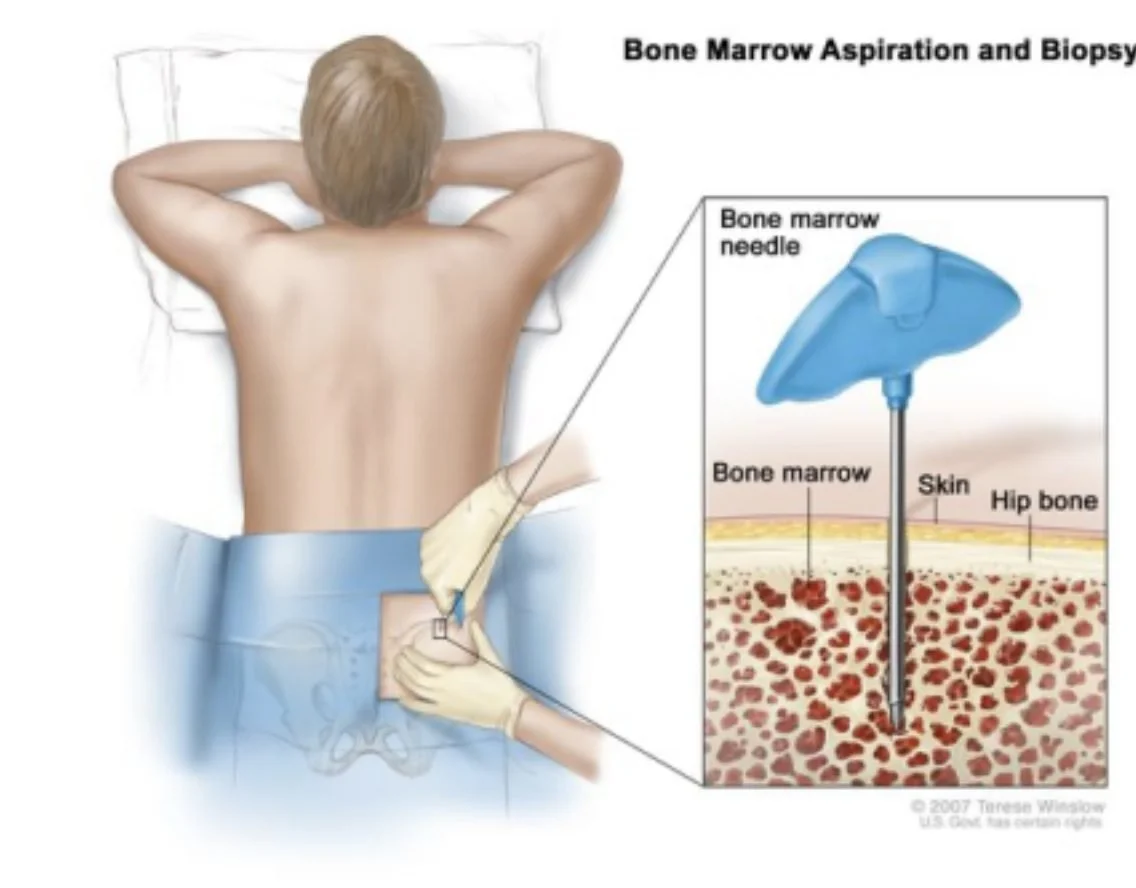
This is normally done in the pelvic bone. Both soft tissue and fluid are removed and then examined by a pathologist. There might also be molecular and genetic tests that can be used to identify the specific subtype of AML. Fluorescence-in-situ-hybridization (FISH) is used to determine if and what chromosomal abnormalities are present.
Figure 4
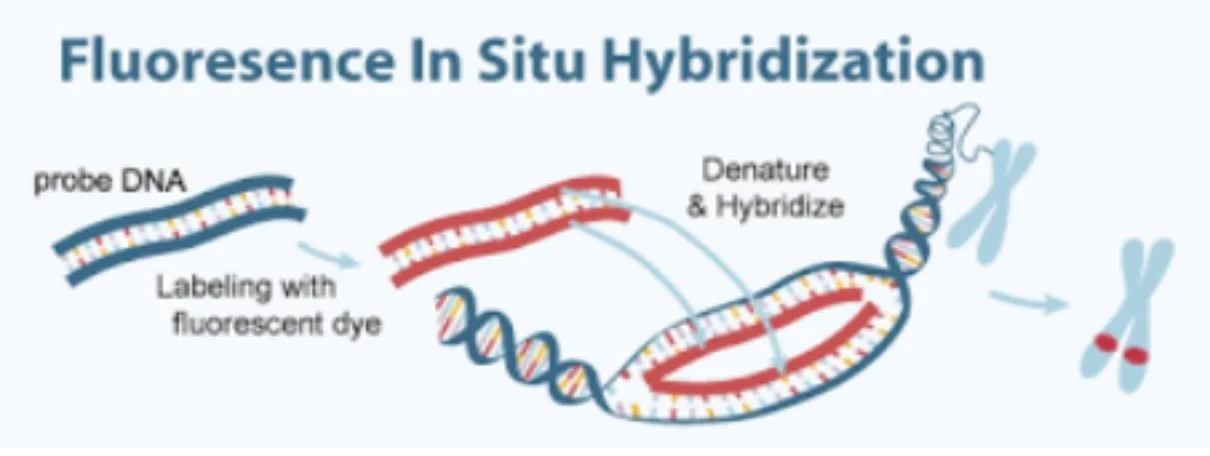
This test is usually conducted on the tissue removed during the bone marrow biopsy or aspiration procedure. It helps the doctors determine the subset of Leukemia. Imaging tests such as a CT, MRI, or a PET scan might also be used to diagnose infections and learn more about the cause of symptoms. A more uncommon diagnostic procedure that can be used is the lumbar puncture (spinal tap). This is used to take a sample of the cerebral spinal fluid to see if there are any AML cells present.
Treatment
Chemotherapy
Chemotherapy is the most useful treatment for treating leukemia because when these anti-cancer drugs are either injected or orally taken, they enter the bloodstream and travel through the entire body. For AML, there are two stages: induction and consolidation. The induction phase typically
only lasts a week and is very intensive, the main goal being to reduce the amount of leukemia cells to normal levels in the bone marrow. Consolidation therapy is given in cycles to destroy any remaining cancer cells in the blood. Patients who are diagnosed with acute promyelocytic leukemia (APL), are given low doses of chemotherapy for months or years after the consolidation phase. The most common chemotherapy drugs used are cytarabine and an anthracycline drug. Some common side effects include hair loss, lack of appetite. diarrhea/constipation, nausea/vomiting, and mouth sores. Because these drugs also affect the normal cells in the bone marrow, there is a higher risk of infections, easy bruising/bleeding, and fatigue/shortness of breath. Oftentimes, drugs are given to increase the white blood cell count. For patients who have an extremely large number of leukemia cells, the induction phase causes tumor lysis syndrome (TLS). The killed cancerous cells release what's inside into the bloodstream, overwhelming the kidneys. This can be prevented by providing the patients with an excess amount of fluid during the chemotherapy.
Targeted Therapy
Patients with AML often have mutations in their FLT3 gene that produces FLT3, a protein that aids the cell's growth. Midostaurin is a drug that blocks FLT3 and other proteins that cause growth in the cancer cells. This drug is taken along with chemotherapy twice a day. Gilteritinib is another drug that inhibits FLT3 and is often given to patients who have had a remission or whose previous treatments have not worked. Another common mutation in patients with AML is in the IDH1 and IDH2 gene. These mutations prevent the blood cells from developing normally. IDH inhibitors such as Ivosidenib (for IDH1 mutation) and Enasidenib (for IDH2 mutation) are given once a day for patients with these mutations. Another targeted therapy is mylotarg which uses a man made monoclonal antibody to attach to a protein called CD33 which is found on AML cells. This antibody works as a signal to alert the chemotherapy drugs to enter and kill the leukemia cell it is attached to. This treatment can be used along with chemotherapy but it is often given to those who are too weak for chemotherapy or for those that are no longer responding to other treatments. Another treatment is venetoclax which works on inhibiting BCL-2 that allows the cancer cells to have an extended life. This is usually given to patients who are 75+ or those who are too weak for strong chemotherapy. Some patients with AML have a mutation in their hedgehog pathway which is needed during embryo and fetus development. Glasdegib targets a protein in this pathway and is given to patients who are older than 75 or not strong enough for chemotherapy. A rare side effect of some of these treatments occurs when the leukemia cells release certain chemicals that can possibly cause liver and kidney damage. This is known as differentiation syndrome. Another possible side effect is TLS.
Surgery
Surgery is rarely used as a form of treatment for AML since the leukemia cells are spread through the blood. If a rare tumor (myeloid sarcoma, chloroma, or granulocytic sarcoma) occurs, it can be removed through surgery. Oftentimes, patients who are diagnosed with AML have a central venous catheter (CVC) placed in their chest to give IV drugs and to take blood samples.
Figure 5
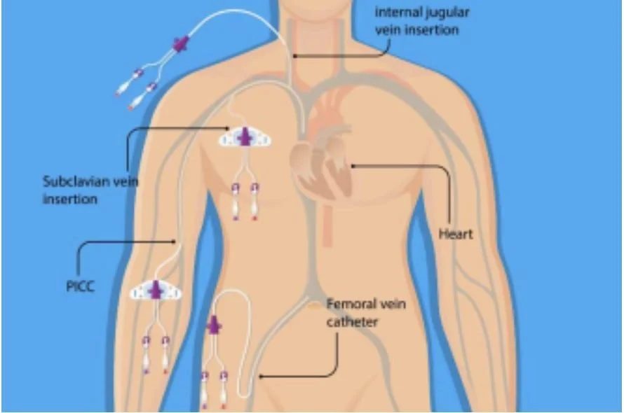
Various catheters are inserted in order to administer IV fluids and take blood samples.
Radiation Therapy
Radiation therapy is a treatment that uses high doses of radiation to kill cancerous cells. Since patients with AML have cancer cells spread throughout their body, it is not commonly used. But it may be used to treat patients whose leukemia has spread outside of the blood such as to the brain and spinal fluid. Before a stem cell transplant, radiation is given to the whole body. Radiation is also used to shrink a tumor although chemotherapy is preferred. It can also be used on bones that are painful if the cancer is not responding to chemotherapy. For AML, external beam radiation is used. Radiation can also cause a decrease in red blood cells, platelets, and white blood cells making the patients more vulnerable.
Stem Cell Transplant
A stem cell transplant (SCT), or a bone marrow transplant, is given when the doctors need to use high doses of chemotherapy for treatment. This damages the patients normal cells leading to dangerous conditions. By giving them a stem cell transplant, they can continue to safely treat the patient. One type of SCT is allogeneic stem cell transplant. This is when the stem cells used come from a person other than the patient. The donor and the patient both have to have the same HLA (tissue) type to avoid an immune response. Autologous stem cell transplant is when the patient’s own stem cells are removed and frozen. While stored, the cells often go through purging to try and remove any leukemia from them. Allogeneic transplant is preferred over this form since the donor will not have any cancerous cells. Sometimes there might be a graft versus leukemia effect in which the donor’s cells see the remaining cancer cells as foreign and attack them. On the other hand, the most severe complication that can arise from a STC is a graft versus host disease in which the donor’s cells take over the patient's immune system and attack the patients tissues. Another possible treatment is non-myeloablative transplant which is when the patient is still given low doses of chemotherapy and radiation that do not completely kill all cancer cells, but is also given a mini transplant of allogenic stem cells. These cells then create a graft versus leukemia effect attacking the remaining cancer.
Treatment Response Rates
2 out of 3 patients with AML that receive standard induction chemotherapy go into remission. The prognosis rates are typically higher for those below 60 while those who are above 60 have lower response rates. A stem cell transplant has a higher rate of success but also has a much higher rate of death as a side effect. 90% of patients with APL go into remission and 80% go into long term remission with proper care. Patients whose AML comes back and does not respond to more intensive chemotherapy are given Mylotarg. If none of the treatments work, the objective shifts towards making the quality of life better, also known as palliative treatment. This would include radiation therapy and strong pain relievers such as morphine. As of now, there are no clear treatments for long term remission in patients that suffer from AML. A recent discovery noted the overexpression of IL1RAP Leukemia stem cells and that decreasing expression of this also decreases the growth and survival of AML cells (Jiang et.al. 2016). They also found that using Lead anti-IL IRAP antibodies would inhibit tumor growth.
Conclusion
AML, acute myelogenous leukemia, is a type of cancer that is formed by stem cells in the bone marrow that do not properly develop into blood cells. Chromosomal errors during DNA replication prevent the cell from developing normally. Patients who have AML tend to be older. There are many different types of AML and treatment can depend on the subtype of AML.
Doctors use blood tests, bone marrow aspirations and biopsies, molecular/genetic tests, FISH, imaging and a spinal tap to diagnose patients. The most common type of treatment is chemotherapy which is given in two stages. This is often accompanied by other targeted therapies which inhibit certain genes or proteins that support the cancerous growth. The overall outlook is positive but depends on the patient. There is still further research being done on how immunotherapy can be used to treat AML. Researchers are trying to use adoptive immunotherapy to increase a patient's white blood cells ability to fight with the tumor. As well as trying to find new ways to treat AML, researchers are also looking to increase the effectiveness of existing treatments and lower the dangerous side effects. This hard work gives hope that patients with AML will soon be able to have better long term remission rates.
References
Albert Einstein College of Medicine. (2017, October 9). Novel treatment causes cancer to self-destruct without affecting healthy cells. ScienceDaily. Retrieved June 19, 2021 from www.sciencedaily.com/releases/2017/10/171009123906.htm
Bernasconi P, Boni M, Cavigliano PM, Calatroni S, Giardini I, Rocca B, Zappatore R, Caresana M, Quarna J. Molecularly targeted therapy in acute myeloid leukemia. Ann N Y Acad Sci. 2004 Dec;1028:409-22. doi: 10.1196/annals.1322.049. PMID: 15650266.
Berenstein R. (2015). Class III Receptor Tyrosine Kinases in Acute Leukemia - Biological Functions and Modern Laboratory Analysis. Biomarker insights, 10(Suppl 3), 1–14. https://doi.org/10.4137/BMI.S22433
Chemotherapy for Acute Myeloid Leukemia (AML). American Cancer Society. (n.d.). https://www.cancer.org/cancer/acute-myeloid-leukemia/treating/chemotherapy.html. Doepfner KT, Boller D, Arcaro A. Targeting receptor tyrosine kinase signaling in acute myeloid leukemia. Crit Rev Oncol Hematol. 2007 Sep;63(3):215-30. doi:
10.1016/j.critrevonc.2007.05.005. Epub 2007 Jul 19. PMID: 17658267.
If Acute Myeloid Leukemia (AML) Doesn't Respond or Comes Back After Treatment. American Cancer Society. (n.d.). https://www.cancer.org/cancer/acute-myeloid-leukemia/treating/recurrence.html.
Leukemia - Acute Myeloid - AML - Subtypes. Cancer.Net. (2017, August 18). https://www.cancer.net/cancer-types/leukemia-acute-myeloid-aml/subtypes. Mayo Foundation for Medical Education and Research. (2021, February 10). Acute myelogenous leukemia. Mayo Clinic.
https://www.mayoclinic.org/diseases-conditions/acute-myelogenous-leukemia/symptoms-c auses/syc-20369109.
Ping Jiang, Bob Y. Liu, Jen Huang, Jennifer Lu, Sharmili Roy, Madhavi Mishra, Xiaoxian Zhao, Jeffrey Lin, Eric D. Hsi and Jagath R. Junutula Cancer Res July 15 2016 (76) (14 Supplement) 3337; DOI: 10.1158/1538-7445.AM2016-3337
Radiation Therapy for Acute Myeloid Leukemia (AML). American Cancer Society. (n.d.). https://www.cancer.org/cancer/acute-myeloid-leukemia/treating/radiation-therapy.html. Risk Factors for Acute Myeloid Leukemia (AML). American Cancer Society. (n.d.). https://www.cancer.org/cancer/acute-myeloid-leukemia/causes-risks-prevention/risk-factor s.html.
Scholl C, Gilliland DG, Fröhling S. Deregulation of signaling pathways in acute myeloid leukemia. Semin Oncol. 2008 Aug;35(4):336-45. doi: 10.1053/j.seminoncol.2008.04.004. PMID: 18692684.
Surgery for Acute Myeloid Leukemia (AML). American Cancer Society. (n.d.). https://www.cancer.org/cancer/acute-myeloid-leukemia/treating/surgery.html. Targeted Therapy Drugs for Acute Myeloid Leukemia (AML). American Cancer Society. (n.d.). https://www.cancer.org/cancer/acute-myeloid-leukemia/treating/targeted-therapy.html.
What Causes Acute Myeloid Leukemia (AML)? American Cancer Society. (n.d.). https://www.cancer.org/cancer/acute-myeloid-leukemia/causes-risks-prevention/what-caus es.html.
What Is Cancer? National Cancer Institute. (n.d.).
https://www.cancer.gov/about-cancer/understanding/what-is-cancer.
