
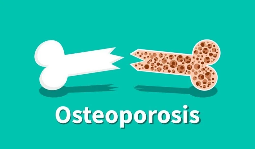
Did you know one in two women over age 50 will break a bone in their lifetime due to osteoporosis? One in two! The chances of developing osteoporosis is greater than the combined risk of heart attack, stroke, and breast cancer, making osteoporosis one of the most important diseases women should be paying attention to. But what is it, and how are we treating it?
Osteoporosis is a disease when the body loses too much bone or makes too much bone, making it easy to fracture bones without any warning signs. By around the age of 30, bones are at peak bone density, and the body will be able to replace whatever bone cells (osteocytes) that die. However, when the bone mineral density is too low, it can turn into osteoporosis, which is most common in people ages 50 and older and with women. Using drugs like glucocorticoids (steroids that lower inflammation), anticonvulsants (used to treat epilepsy by blocking the GABA neurotransmitter), heparin (medication used as an anticoagulant during heart attacks), and alcohol may increase the risk of developing osteoporosis.
Osteoporosis does not show symptoms until you break a bone -- meaning parents may not know they have weak bones until they end up in the hospital unexpectedly. As a result, osteoporosis is called “the silent disease.” In order to prevent one from having adverse complications due to osteoporosis, it is essential to notify your parents to get a bone density test to spot bone loss before it starts showing symptoms. Bone density tests are now used by DEXA scans, or dual-energy X-ray absorptiometry scans. These are painless tests that take perhaps 5 minutes to scan a person with minimal radiation -- the radiation using a DEXA scan is almost undetected by standard radiation monitors and are better and faster than X-rays.
Current treatments and research for osteoporosis is limited to mostly ingesting calcium and maintaining a healthy lifestyle. Medications include receptor activator of nuclear factor kappa-B ligand inhibitors (RANKL), bisphosphonates, estrogen agonists/antagonists, parathyroid hormone analogues, and calcitonin (Tu et al., 2018).
Osteoclastogenesis is the process of breaking down bones, where osteoclasts are osteocytes found on the surface of the bone which are undergoing resorption (the breaking down of bone cells). They clump together to create resorption bays, where they secrete acid and collagenase to break down bone cells (Yavropoulou & Yovos, 2008). They have a ruffled border where it anchors onto surrounding bone cells, secreting hydrogen ions, collagenase, cathepsin K, and hydrolytic enzymes. This zone is where the osteoclast will dissolve minerals and organic materials in what is called the “clear zone” or “sealing zone.” Because osteoclasts basically ingest bad bone cells, they are considered to be a part of the mononuclear phagocyte system, and they are regulated by hormones (chemical signals made by specific glands) and cytokines (other chemical signals, but they are made from immune cells). In other research, knowledge of specific cytokines is essential for creating new therapies for osteoporosis and other diseases.
An osteoblast is a cell with a singular nucleus that helps create other osteocytes. Osteoblasts must coagulate into a group of cells called an osteon in order to make bones. Together, osteons create bone through one of two processes: endochondral ossification or intramembranous ossification. Endochondral ossification is most common, where chondrocytes create cartilage using collagen and proteoglycans. Your ears, skin, and nose are all made of cartilage, but what makes them differently textured is the ratio between collagen and proteoglycans. Proteoglycans are the “filler” substance between cells, while collagen gives the rigid structure to your ear or bridge of your nose. Together, cartilage is grown through interstitial growth and can be transformed into bone through a process called ossification.
The cartilage-to-be-bone is called the perichondrium. Ossification starts in the center of the bone, where osteoprogenitor cells (undifferentiated cells) become osteoblasts, creating the periosteum. Afterward, the osteoblasts form the bone collar, which is the shape of how the new bone is going to grow. Afterwards, the bone collar makes a sort of scaffold in which chondrocytes begin secreting alkaline phosphatase, an enzyme used in mineral deposition. There, osteoblasts secrete osteoid which forms the bone trabeculae, the calcium-filled spongy tissue in the middle of the bone which is broken down into bone marrow.
Once mammals are born, second ossification is the process in which your bone grows longer. Periosteal buds are groups of embryonic stem cells called mesenchyme that invade the bone when you are born, rich in cells that will differentiate into blood, bone, and other cells necessary to grow. This part in which your bones grow is separated by the epiphyseal plate where bone is grown from the center versus the ends of the bone. It continues to increase in length until an individual is about 20 years old where we reach peak bone growth and until 30 years when we reach peak bone density.
Intramembranous ossification is the second type of way the bones develop in the fetus, and it does not require collagen to produce bone (Zhang et al., 2018). Mesenchymal stem cells instead transported by periosteal buds carry cells with large, round nuclei that are surrounded by chromatin, making these cells appear clear. They have a small amount of Golgi apparatus, rough endoplasmic reticulum, mitochondria, and ribosomes. The process of ossification starts with bone cells starting to replicate in a small cluster of cells called a nidus. Once the nidus is created, all the cells start developing into osteoprogenitor cells which will differentiate into osteoblasts. They then create type-I collagen fibrils which stimulate the development of bone tissue and mineralization in the bone.
Receptive activators of the nuclear factor kappa-B ligand inhibitors (RANKL) is a tumor necrosis factor, meaning it breaks down cells that no longer work. It is important in binding to the receptor RANK that is on the surface of osteoclasts, osteoblasts, and other bone cells. They help activate osteoclasts to start breaking down bone and remodeling them, but they also prevent osteoclasts from apoptosis, making them cancerous if they are not regulated by osteoprotegerin (OPG) which competitively inhibits RANKL and can be used as a medicine in fighting osteosarcomas. Among other causes for bone cancer include gene mutations, parathyroid hormone (regulating calcium and phosphorous in the body), vascular endothelial factor (VEGF, angiogenic factor which helps in bone formation, blood formation, and wound healing), and interferons (cytokines produced by cells infected by a virus which signal other cells to stay clear or to invite immune cells to come).
Bisphosphonates are drugs that are similar to the chemical inorganic pyrophosphate (PPi) that is made by the body in blood and urine. It is capable of inhibiting calcification, which is important because those who suffer from osteoporosis have their bones overly calcified, leading to brittle bones. Recently, some research suggests that bisphosphonates limit apoptosis, which may become cancerous if used excessively (Plotkin, 2005).
More research is needed to know whether current treatments for osteoporosis actually work since they only slow down the breaking down of bones. Bone regeneration would be the most efficient way to manage osteoporosis, but what is the current research, and how can one be careful of bone cancer? As our generation continues to age, it becomes crucial to continue researching and learning about the chemical pathways that affect the breakdown and regeneration of bone.
Most of all, those holding Medicare will be able to schedule a bone density test every 2 years. Schedule now by searching up on Medicare’s website and ask your doctor for you or your parents.
Citations
Luo J, Sun MH, Kang Q, Peng Y, Jiang W, Luu HH, Luo Q, Park JY, Li Y, Haydon RC, He TC. Gene therapy for bone regeneration. Curr Gene Ther. 2005 Apr;5(2):167-79. doi: 10.2174/1566523053544218. PMID: 15853725.
Plotkin LI, Aguirre JI, Kousteni S, Manolagas SC, Bellido T. Bisphosphonates and estrogens inhibit osteocyte apoptosis via distinct molecular mechanisms downstream of extracellular signal-regulated kinase activation. J Biol Chem. 2005 Feb 25; 280(8):7317-25.
Tu, K. N., Lie, J. D., Wan, C., Cameron, M., Austel, A. G., Nguyen, J. K., Van, K., & Hyun, D. (2018). Osteoporosis: A Review of Treatment Options. P & T : a peer-reviewed journal for formulary management, 43(2), 92–104.
Yavropoulou MP, Yovos JG. Osteoclastogenesis--current knowledge and future perspectives. J Musculoskelet Neuronal Interact. 2008 Jul-Sep;8(3):204-16. PMID: 18799853.
Zhang, H., Shi, X., Wang, L., Li, X., Zheng, C., Gao, B., Xu, X., Lin, X., Wang, J., Lin, Y., Shi, J., Huang, Q., Luo, Z., & Yang, L. (2018). Intramembranous ossification and endochondral ossification are impaired differently between glucocorticoid-induced osteoporosis and estrogen deficiency-induced osteoporosis. Scientific reports, 8(1), 3867. https://doi.org/10.1038/s41598-018-22095-1
