
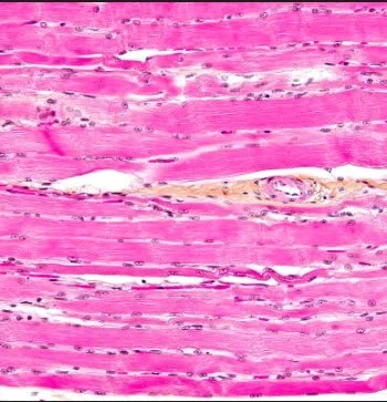
There are three types of muscle tissue: cardiac, smooth, and skeletal (also called striated muscle). Skeletal muscle is unique among these three because it is the only muscle that is under voluntary control. When a person decides to move, it is their skeletal muscle that is responsible for carrying out that action. The movements a person makes is a result of their skeletal muscles contracting.
Skeletal muscle cells are, at a basic level, composed of the protein filaments actin and myosin. Myosin, or the thick filament, is about 15nm in diameter and provides the “power stroke” of muscle contraction. It has been dubbed a molecular motor, and also acts as an ATPase enzyme. Actin, or the thin filament, is about 7 nm in diameter. It has two important proteins attached to it that will be discussed later. When actin forms the thin filament in skeletal muscle cells, it has a helical shape: in this case, the actin filament is composed of two chains of actin molecules twisting around each other.
Muscle contraction begins with the release of the neurotransmitter acetylcholine (ACh). This begins when an upper motor neuron sends a signal to a lower motor neuron to contract. This signal propagates to the axon terminal of the LMN and causes an influx of calcium ions to the axon terminals from voltage-gated channels. There are vesicles in the axon terminal that contain ACh, which have proteins on the outside that will bind with the calcium ions. Once the calcium binds the vesicle, the entire thing is drawn to the membrane of the axon because of the differing charges. Once the vesicle reaches the membrane, it will fuse with it to become one continuous membrane. This releases the contents of the vesicle, the ACh, into the synaptic cleft in a form of exocytosis.
This synapse, between the muscle cell and the motor neuron, is called the neuromuscular junction.
Once in the synaptic cleft, the ACh molecules are able to bind to receptors, called nicotinic acetylcholine receptors (nAChR), located on sodium channels of the postsynaptic muscle cell. When ACh binds to the nAChRs, the sodium channel receives a signal to open. An influx of sodium cations flow into the muscle cell, which depolarizes it. As the influx of Na2+ into the cell is dependent on the voltage of the membrane, this is called voltage-dependent release. These sodium cations turn the membrane potential positive. Voltage-gated channels activate with this change in potential, causing an action potential throughout the muscle cell.
Transverse (or “T”) tubules criss cross the cell, and spread the action potential (the depolarization) throughout it. This action potential activates L-type Ca2+ channels (a type of voltage-dependent calcium channel). These channels are mechanically coupled to the sarcoplasmic reticulum ryanodine receptors. The sarcoplasmic reticulum is an organelle that contains Ca2+. Ryanodine receptors (RyRs) are the main Ca2+ release channels for the SR. The L-type Ca2+ channels modulate the RyRs, and therefore modulate the release of Ca2+.
Once the action potential has activated the L-type Ca2+ channels, this mechanical coupling prompts the release of Ca2+ from the sarcoplasmic reticulum into the muscular cytoplasm. Now that Ca2+ is available for usage in the cell, contraction can begin.
With the newly present Ca2+ begins a series of movements within the muscle cell that leads to contraction.
As previously described, there are two important proteins associated with the actin filament. One of these proteins is called tropomyosin. It is a protein that wraps around the actin filament, and is secured to the actin by the protein troponin. There are Ca2+ binding sites on the troponin, and when Ca2+ is present, like it is after release from the sarcoplasmic reticulum, Ca2+ binds to the troponin. Ca2+ binding to troponin causes the tropomyosin to shift position and expose previously covered sites on the actin filament. These sites are known as myosin-binding sites, because those sites are where the myosin heads bind during muscle contraction.
It takes energy for the myosin “head” to cock into a higher energy position, so that it might bind with the actin. The energy used is ATP, which is the energy biological systems use.
One starting point for this situation is as follows: the myosin head is attached to its place on the actin, in a low energy position, and not able to move up the actin (contraction). This configuration, where the myosin head attached to the actin filament, is called a cross bridge.
Contraction can begin when ATP is made available to the myosin, and it binds to the myosin head. This gives the myosin the required burst of energy to release the actin filament. Once the myosin head is no longer attached to the actin, it begins to hydrolyze the ATP attached to it. It does this because it is an ATPase, an enzyme that acts as a catalyst in the hydrolysis of ATP. ATP hydrolysis involves the ATP molecule getting a phosphate pulled off of it, resulting in an ADP molecule, an inorganic phosphate, and energy release. This energy allows for the myosin head to “cock” into a higher energy state, changing its conformation. When that free phosphate is released from the myosin, the myosin receives an additional energy burst. This is enough for the “power stroke,” where the myosin reattaches to the actin (forming another cross bridge), and pulls on it. This is contraction.
This process is known as “sliding filament theory.” The idea behind sliding filament theory is that actin and myosin “slide” past each other, forming cross bridges, to create muscle contractions.
The reason the actin doesn’t “slide back” after the myosin head has released it is because there are many myosin heads all going through the same process at the same time, so that there are always some attached to the actin and some cocking back and creating the power stroke.
As myosin heads pull on actin filaments, the muscle fiber “bunches up” or “shortens.” Eventually, when the myosin heads cannot stretch any further, the fiber will reach a point when it can no longer continue contraction.
Muscular relaxation begins once stimulation of the motor neuron no longer occurs. When ACh is no longer being delivered, Na2+ is no longer entering and depolarizing the fiber membrane, and as action potentials are no longer being triggered, Ca2+ is no longer released from the sarcoplasmic reticulum. The remaining Ca2+ in the cell is pumped back into the sarcoplasmic reticulum, and acetylcholinesterase destroys the remaining ACh left in the synaptic cleft.
Research in the area of muscle contraction has led to some fruitful results. With the knowledge of how muscle contraction happens, scientists are able to better design medicine, cures, and therapies for people with muscular disorders.
Word Bank:
-- Helical: in the shape of a helix (think of DNA)
-- Propagate: to spread around
-- Vesicle: a very small pocket that contains molecules of specific neurotransmitters
-- ATP hydrolysis: the breaking down of ATP molecules in order to release energy
-- Voltage-gated/dependent ion channel: an ion channel that responds to changes in electrical stimuli
Sources:
Aaronson, Kim, and et al. “10.3 Muscle Fiber Contraction and Relaxation.” Anatomy and Physiology, OpenStax, 6 Mar. 2013, opentextbc.ca/anatomyandphysiology/chapter/10-3-muscle-fiber-contraction-and-relaxation/.
Cooper, Geoffrey M. “Actin, Myosin, and Cell Movement.” The Cell: A Molecular Approach. 2nd Edition., U.S. National Library of Medicine, 1 Jan. 1970, www.ncbi.nlm.nih.gov/books/NBK9961/.
Kuo, Ivana Y, and Barbara E Ehrlich. “Signaling in Muscle Contraction.” Cold Spring Harbor Perspectives in Biology, Cold Spring Harbor Laboratory Press, 2 Feb. 2015, www.ncbi.nlm.nih.gov/pmc/articles/PMC4315934/.
Lanner, Johanna T, et al. “Ryanodine Receptors: Structure, Expression, Molecular Details, and Function in Calcium Release.” Cold Spring Harbor Perspectives in Biology, Cold Spring Harbor Laboratory Press, Nov. 2010, www.ncbi.nlm.nih.gov/pmc/articles/PMC2964179/.
Nature News, Nature Publishing Group, www.nature.com/scitable/content/troponin-and-tropomyosin-regulate-contraction-via-calcium-14723328/.
Schmidt, William M, et al. “Direct Observation of Tropomyosin Binding to Actin Filaments.” Cytoskeleton (Hoboken, N.J.), U.S. National Library of Medicine, June 2015, www.ncbi.nlm.nih.gov/pmc/articles/PMC4529784/.
Walker, Warren F., and Norman R. Alpert. “Muscle Types.” Encyclopædia Britannica, Encyclopædia Britannica, Inc., 8 Aug. 2019, www.britannica.com/science/muscle/Muscle-types#ref58933.
Image Credit: https://upload.wikimedia.org/wikipedia/commons/thumb/3/38/Skeletal_striated_muscle.jpg/1200px-Skeletal_striated_muscle.jpg
Teens In Health Student Submission Winner
