
The Discovery of X-Rays
Have you ever seen x-rays on the TV, where you could see someone’s bone right through a screen? Surprisingly, x-rays have been discovered and used for medical practice for over 100 years now! X-rays are also called Röntgen radiation, after the German professor Wilhelm Röntgen who discovered them. In the 1800s, some scientists who were studying cathode rays came across with another high-energy ray. (These rays would eventually lead to the discovery of electrons.) When Röntgen was experimenting with these cathode rays in a special Crookes Tube, a glass tube used to examine cathode rays, he noticed that another ray was capable of passing through books, emitting a light that cast a shadow. These rays, which he named “X-ray” for being a variable ray, could also pass through a person’s hand, only emitting the shadow of their bones. By 1895, Röntgen submitted his discovery and won the first Nobel Prize in Physics (Harisinghani et al., 2018).
With the discovery of X-rays came many inventions in science. From removing bullets to analyzing fractured bones, many doctors could now have a way to see their patient from the inside without ripping them apart in surgery. During World War I, Marie Curie used X-rays to build cars to help screen injured soldiers. X-rays were also found to be able to be diffracted using crystals, which led to the discovery of X-ray crystallography. This technique would be used by James Watson, Francis Crick, Maurice Wilkins, and Rosalind Franklin to discover the helical structure of DNA that we know today. Much in the future, South African American physicist Allan M. Cormack and British electrical engineer Godfrey N. Hounsfield invented the computed tomography scan (commonly known as the CT scan), which uses multiple x-rays in order to create images of cross-sections of a body part. This would become immensely helpful for researching cross-sections of the brain, heart, and lungs (Garvey & Hanlon, 2002).
X-Rays Drawbacks
However, x-rays had their setbacks. X-rays, like all types of light, are caused by photons moving at different wavelengths. X-rays have particularly short wavelengths, meaning they’re protons that move incredibly quickly. As a result, x-rays have much more energy than the light we can see, and this means they have the power to damage human cells, causing radiation sickness, developmental issues, and cancer (De Santis et al., 2007). CT scans also require using contrast agents, which are chemicals that make images clearer. When a person takes these chemicals, they run a risk for poisoning. As a result, contemporary radiology have come up with more inventions that minimize the amount of radiation caused to the patient as well as the healthcare workers that deal with them, such as the DXA scan.
The DXA scan
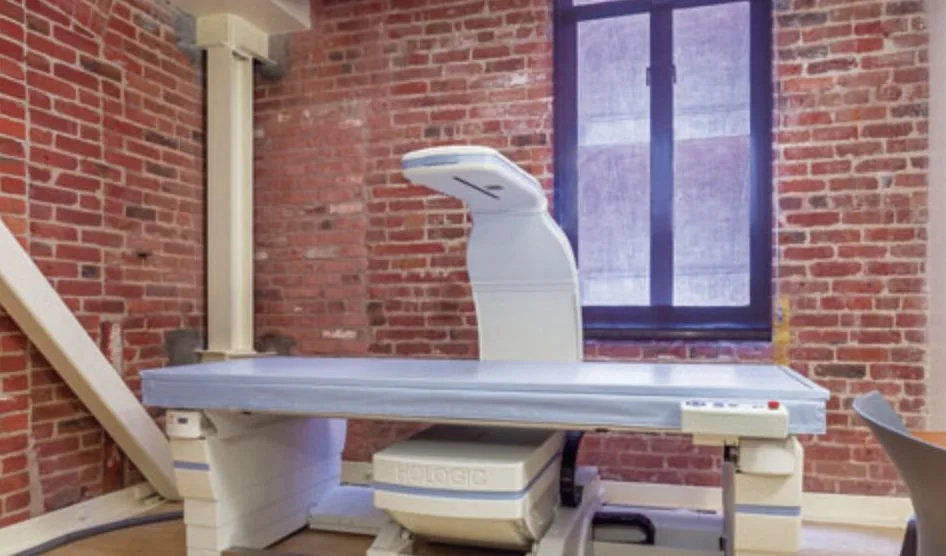
The DXA, DEXA, or dual-energy X-ray absorptiometry uses two different types of x-rays of different wavelengths in order to produce pictures of a person’s skeleton as well as their body fat and muscular composition. Soft tissue and bone have different “attenuation coefficients,” which are numbers that measure how much they reflect or absorb x-rays. Meaning, x-rays will pass through soft tissue while not for bone (which is why we can see shadows for bones on an x-ray). By using two different x-rays of different wavelengths, these x-rays will go through different parts of the body and create a double-layered image to tell a radiologist the mineral, fat, and muscular composition of a patient. Using a DXA scan, the images are scanned by a computer to calculate the mineral content in the bone in relation to a healthy counterpart using the Z score and compared to a young adult using the T score. These calculations are then able to determine whether a person is at risk for issues such as osteoporosis, when the bone has a low mineral density (Blake & Fogelman, 2007).
Effects of DXA and New Inventions
With DXA scans, many more people are able to be diagnosed and assisted earlier. Osteoporosis results in an increased risk of fracture, which makes it important to note when around 54 million people have low bone density in the United States, which is about 1 in 6 people. As a result, it becomes necessary for people, especially those who are 50 years old and older, to receive periodic checkups on their bones to decrease their risk and prevent it using lifestyle and diet interventions, such as weightlifting (which can train bones to hold more weight) and eating calcium and vitamin D-fortified foods (which can help bones to grow, for kids, and maintain, for adults). Modern DXA scans now emit a level of radiation that is undetectable by radiation detectors (Bushberg et al., 2002). Moreover, with more inventions for medical uses, the cost of an X-ray has now decreased significantly. Now, inventions such as MRI (Magnetic Resonance Imaging) help doctors even see soft tissue and brain matter more easily, not just bones (Grover et al., 2015). This allows doctors to catch inflammation and cancer much more easily than CT or other X-ray scans. From their discovery, X-rays have shown to be pivotal in diagnosing many different types of illnesses. Although they had their setbacks, modern-day technology works on improving their efficiency and reducing their risk. It becomes the question now to how imaging technology will be able to be accessible for the general public, and not just modern day, and how they can be possibly used in other ways, such as imaging chemicals in food or contaminated water.
Citations
Blake, G. M., & Fogelman, I. (2007). The role of DXA bone density scans in the diagnosis and treatment of osteoporosis. Postgraduate medical journal, 83(982), 509–517. https://doi.org/10.1136/pgmj.2007.057505
Bushberg, Jerrold T.; Seibert, J. Anthony; Leidholdt, Edwin M.; Boone, John M. (2002). The essential physics of medical imaging. Lippincott Williams & Wilkins. p. 38. ISBN 978-0-683-30118-2.
De Santis M, Cesari E, Nobili E, Straface G, Cavaliere AF, Caruso A (2007). "Radiation effects on development". Birth Defects Res. C Embryo Today. 81 (3): 177–82. doi:10.1002/bdrc.20099. PMID 17963274.
Garvey, C. J., & Hanlon, R. (2002). Computed tomography in clinical practice. BMJ (Clinical research ed.), 324(7345), 1077–1080. https://doi.org/10.1136/bmj.324.7345.1077
Grover, V. P., Tognarelli, J. M., Crossey, M. M., Cox, I. J., Taylor-Robinson, S. D., & McPhail, M. J. (2015). Magnetic Resonance Imaging: Principles and Techniques: Lessons for Clinicians. Journal of clinical and experimental hepatology, 5(3), 246–255. https://doi.org/10.1016/j.jceh.2015.08.001
Harisinghani, Mukresh G., Chen, John W., Weissleder, Ralph. Primer of diagnostic imaging. 2018. ISBN : 9780323377508
Image Credit to UCSF Radiology Center: https://radiology.ucsf.edu/patient-care/services/bone-density-scan-dxa-dexa
